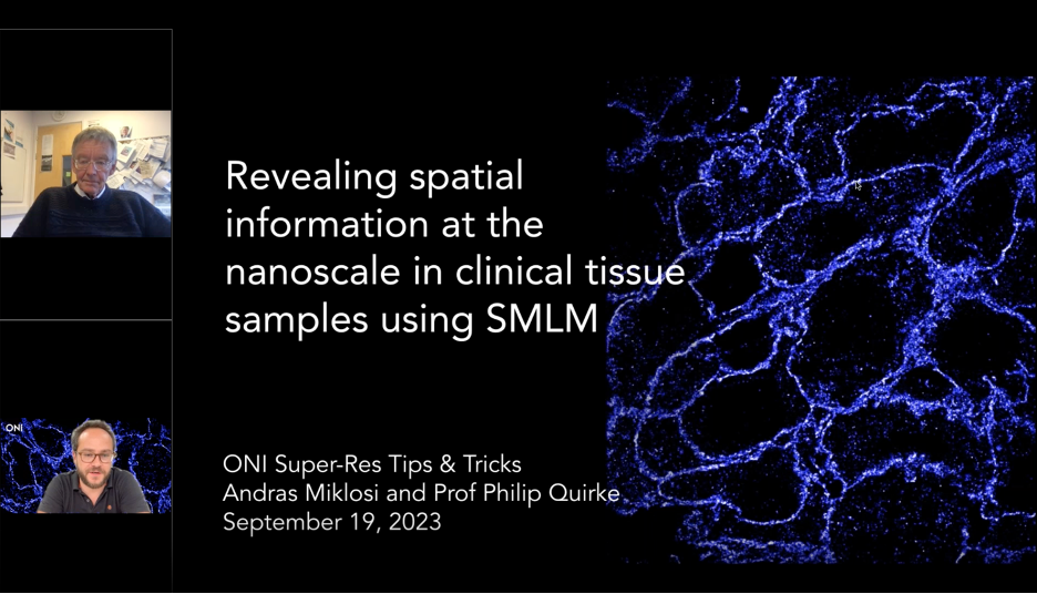
Single-Molecule Localization Microscopy (SMLM) offers high sensitivity and resolution, which can enable the detection and quantification of spatial information across a range of tissue samples. Information about the underlying physiology of nanoscale structure reorganization often bears clinical relevance and, as such, SMLM microscopy is becoming an increasingly popular tool in pathology and diagnostic imaging.
This Tips & Tricks webinar was presented by ONI’s Senior Scientist Andras Miklosi, in partnership with Prof. Philip Quirke from the University of Leeds. The webinar covers tips & tricks around sample preparation, imaging and analysis, with a focus on how SMLM is being applied across a range of applications, including protein targets associated with tumor biology. The clinical potential of super-resolution microscopy for renal pathology diagnostics is emphasized through data from the ongoing collaboration between ONI and the University of Leeds, which is funded by the NIHR i4i programme.

To discover more about the super-resolution microscopy and how our Nanoimager can be used, visit our ONIversity.